MCC-CC Listings for MS-DRGs FY2024

The CMS FY2024 Final Rule tables include: Complete MCC List; Complete CC List; Additions to the MCC List; Deletions to the MCC List; Additions to the CC List; Deletions to the CC List.

To download these listings, CLICK HERE.

Get our CDI Pocket Guide® to learn more about important MCCs and CCs that impact MS-DRGs and APR-DRGs.

PEPPER Report: Important Resource to Measure How You're Doing

Your PEPPER report is an excellent (and free) source of data into areas of potential coding risk and specifically to provide guidance on where to focus your hospital’s auditing and monitoring efforts.  It is always better to know if you have a potential problem so that corrective action can be taken before the auditors come knocking.

What is the PEPPER Report?

PEPPER is an acronym for Program for Evaluating Payment Patterns Electronic Report. It is a report that provides provider-specific traditional Medicare data statistics for discharges or services at risk for improper payments. PEPPER is developed and distributed quarterly by TMF Health Quality Institute under contract with CMS.

PEPPER should be used as a guide to identify possible payment errors that includes both over-coding and under-coding, as well as medical necessity issues.  CMS audit contractors may focus on similar target areas as those included in PEPPER.

Each PEPPER contains hospital-specific statistics for the most recent 12 federal fiscal quarters for each target area that may indicate a risk for improper payments.

Understanding Outliers

Outliers are findings that differ significantly from what most organizations are reporting.  Because they lie ‘outside’ the normal bell curve of findings, outliers can be used to identify organizations that may be over-coding or under-coding their cases.

PEPPER flags outliers as the 80th percentile and 20th percentile of all hospitals nationally, jurisdiction (MAC/FI), and state. Each quarter, all the target areas are listed on the “Compare” worksheet with the numerator (“Number of target discharges”) and the numerator and denominator ratio (expressed as “Percent”) along with comparative national, jurisdictional, and state percentiles.

If the ratio (“Percent”) is a jurisdictional outlier it is flagged as follows:

Example of Outliers

Let’s look at an example for two of the PEPPER target areas: Medical DRGs with CC or MCC and Surgical DRGs with CC or MCC:

For the hospital in this example, the percent of all medical DRGs that include an CC or MCC is 60.2%. For Surgical DRGs, 63.8% of the surgical DRGs have a CC or MCC.  How do we know if these percents are low or high? Good or bad? 

The percentiles help with this by providing a comparison of the hospital’s rate to all hospitals nationwide and by jurisdiction and state.

For Medical DRGs with CC or MCC, compared to all hospitals nationally, the hospital’s 60.2% for medical DRGs is in the 50th percentile (rounded from 50.3). 

What that means is 50% of all hospitals have a higher Medical DRGs with CC or MCC rate than this hospital’s rate, which would be considered average.  Because the 50th percentile is not in the 20th or 80th percentile, it is not noted in green or red.

This hospital’s Surgical DRGs with CC or MCC rate however is 63.8% and is reported in red.  When compared to all hospitals nationally, this hospital’s rate is in the 83rd percentile nationally. That means that 83% of all hospitals have a lower Surgical DRGs with CC or MCC rate than this hospital. Because it is above 80th percentile, it is noted in red.

PEPPER percentiles at or above the 80th percentile (high outlier) for any target area may indicate that the hospital is at a higher risk for improper Medicare payments, i.e., over-coding. The greater the percentile value, particularly the national and/or jurisdiction percentile, the greater the consideration should be given to that target area.

Get our CDI Pocket Guide® for more information regarding accurate coding and documentation of PEPPER target areas.    

Using the PEPPER Report

So how should this hospital in our example identify if there is possibly “over-coding” of MCC/CCs for their surgical cases?  Included in the target stats above is the total number of target discharges that have an MCC or CC (numerator).  In addition to these statistics, PEPPER also provides a detailed listing of these 150 discharges.  By reviewing these 150 cases or a statistically valid percentage of these cases, the hospital can determine if there truly is a risk of over-coding, or this percentage is accurate and supported for their hospital. If over-coding (or under-coding) issues are found, corrective action can be taken.  

The HIM department should become familiar with and routinely review their quarterly PEPPER report to help identify opportunities for improvement and guide their audit and compliance activities. If you haven’t been provided with this report, contact your hospital’s quality department who can download PEPPER from the PEPPER resources portal.  

Note: There has been a temporary pause in distributing PEPPERs through the fall of 2024 as CMS works to improve and update the program and reporting system.  While your organization’s older PEPPER reports can be used to identify general areas of concern, up-to-date findings are not now available.

New Pediatric Sepsis Definition and Criteria

Since 2005, the diagnosis of pediatric sepsis has been based on the pediatric sepsis definitions and criteria of the 2005 International Pediatric Sepsis Consensus Conference: Definitions for Sepsis and Organ Dysfunction in Pediatrics published in Pediatric Critical Care Medicine. These were based on pediatric SIRS criteria.

A new International Consensus Criteria for Pediatric Sepsis and Septic Shock was published in March 2024 in JAMA which is now based on the Phoenix Sepsis Score. There are many similarities to the 2016 Sepsis-3 criteria for the adult population. For Sepsis-3, sepsis is defined as a suspected/confirmed infection with life-threatening organ dysfunction based on six different organ systems as determined by the SOFA score of at least 2 points. 

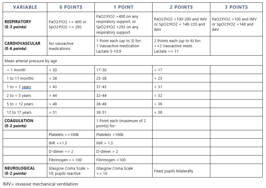
Sepsis in children is now identified by a Phoenix Sepsis Score of at least 2 points in children with suspected/confirmed infection, which indicates potentially life-threatening dysfunction of four organ systems: respiratory, cardiovascular, coagulation, and/or neurological. Septic shock is defined as meeting sepsis criteria with any cardiovascular dysfunction indicated by at least 1 or more points from the cardiovascular organ system.

The new pediatric sepsis definition (like Sepsis-3) requires acute organ dysfunction as part of the definition of sepsis.  Therefore, all cases which meet the pediatric sepsis definition would meet the definition of severe sepsis.  

Phoenix Sepsis Scoring System:

For example, a 6-year-old child with a PaO2/FIO2 (P/F ratio) < 400 on oxygen and on vasoactive medications for hypotension would meet the criteria for septic shock.

Find the complete Pediatric Sepsis definitions and criteria in the CDI Pocket Guide® Unbound Edition.

What can we do?

The new International Consensus Criteria for pediatric sepsis states that the former criteria based on systemic inflammatory response syndrome (SIRS) should no longer be used to diagnose sepsis in children. It’s important that hospitals educate their medical staff on this new definition. 

CDI and coding professionals should begin using the new pediatric sepsis definitions and Phoenix Sepsis Score for clinical validation. Payer auditors may start using these new definitions and criteria for the clinical validation of pediatric sepsis. Successful appeal is unlikely unless Phoenix Sepsis Score criteria are met. Payers should not apply these criteria retroactively to discharges prior to March 2024.


Pinson & Tang has recently partnered with Pediatric Resource Group (PRG) to provide comprehensive pediatric and neonatal content within the CDI Pocket Guide® Unbound Edition that includes over 40 pediatric key reference topics. This collaboration with PRG marks a significant milestone in Pinson & Tang's mission to provide cutting-edge resources for healthcare professionals.

Acute Respiratory Failure - All There Is To Know

The diagnosis and documentation of respiratory failure continues to be challenging for coders, documentation specialists, and physicians. Many physicians, including some intensivists and pulmonologists, are unaware of the widely recognized clinical standards for diagnosing acute respiratory failure even though multiple clinical criteria and appropriate management of respiratory failure are often clearly documented in the medical record. In this article, we will discuss a variety of clinical indicators and tools to identify respiratory failure.

Acute respiratory failure is classified as hypoxemic (low arterial oxygen levels), hypercapnic (elevated levels of carbon dioxide gas), or a combination of the two. In most cases one or the other predominates.

Most patients with acute respiratory failure demonstrate either impaired ventilation or impaired oxygen exchange in the lung alveoli.

In some cases, like head trauma, drug overdose, or over-sedation, the brain’s respiratory center is suppressed causing reduced respiratory drive with decreased ventilation that may progress to respiratory arrest.

For a patient to have acute respiratory failure, it must be symptomatic and meet diagnostic criteria based on arterial blood gas (ABG) or pulse oximetry readings (SpO2).

Get our CDI Pocket Guide® for more information about acute and chronic respiratory failure.    


Blood Gas Measurements

Arterial blood gas (ABG) and pulse oximetry (SpO2) are two methods of measuring blood gases.  Let’s first define what they are:

Measure Definition Normal
pO2 Partial pressure of oxygen, or oxygen content, in mmHg pO2 > 80 mmHg
pCO2 Partial pressure of carbon dioxide, or carbon dioxide content, in mmHg 35 – 45 mmHg
pH Measure of the degree of acidity 7.35 – 7.45
SaO2 Oxygen saturation (percent of hemoglobin carrying oxygen) as reported on ABG and is relatively proportional with pO2 > 95%
SpO2 Oxygen saturation (percent of hemoglobin carrying oxygen) as measured by pulse oximetry and is relatively proportional with pO2 > 95%
FIO2 Percent of supplemental oxygen expressed as a decimal, e.g., 40% oxygen = 0.40. Room air = 20% = 0.20
P/F ratio pO2 / FIO2 > 400

Next, what are the degrees of hypoxemia based on pO2 and/or SpO2 measured on room air, or the P/F ratio measured on supplemental oxygen.

Measure Normal Hypoxemia Acute respiratory failure
pO2 (room air) > 80 mmHg 60-80 mmHg < 60 mmHg
SpO2 (room air) > 95% 91-95% < 91%
P/F Ratio (on oxygen) > 400 300-400 < 300

Hypoxemic Respiratory Failure

Diagnostic criteria:

ImportantP/F ratio and room air pO2/SpO2 criteria are not valid with oxygen-dependent chronic hypoxemic respiratory failure, but pO2/SpO2 criteria are valid for such patients when measured while breathing their usual home O2 flow rate or higher. 

The gold standard for the diagnosis of acute hypoxemic respiratory failure is an arterial pO2 on room air less than 60 mmHg measured by arterial blood gases (ABG). In the absence of an ABG, SpO2 less than 91% measured by pulse oximetry on room air can serve as a substitute for the pO2 because SpO2 of 91% equals pO2 of 60 mmHg.

These criteria may not apply to patients with chronic hypoxemic respiratory failure (e.g., due to severe COPD), because their room air pO2 would always be expected to be < 60 mmHg (SpO2 < 91%).  Chronic respiratory failure patients only qualify for home oxygen when their SpO2 is less than 91% and they are treated with supplemental oxygen on a continuous outpatient basis that is adjusted to keep arterial oxygen saturation above 92%.


Hypercapnic Respiratory Failure

Diagnostic criteria:

*A normal pH of 7.35-7.45 indicates chronic hypercapnic respiratory failure only.

The hallmark of acute hypercapnic respiratory failure is elevated pCO2 due to acute retention/accumulation of carbon dioxide gas resulting in an acidic pH less than 7.35. There are many causes, but severe COPD is the most common.

Physicians may identify these findings as “respiratory acidosis,” which is the same thing as hypercapnic respiratory failure. Respiratory acidosis and acute respiratory acidosis are indexed as hypercapnic respiratory failure and acute hypercapnic respiratory failure, respectively.

Finally, an exacerbation of symptoms requiring an increase in chronic supplemental oxygen indicates an “acute exacerbation” of chronic respiratory failure, which would be classified as acute-on-chronic respiratory failure if properly documented.

With the exception of P/F ratio, Coding Clinic, Third Quarter 1988, p. 7 and Second Quarter 1990, p. 20 have identified the above diagnostic criteria for acute hypoxemic and hypercapnic respiratory failure to assist coders and documentation specialists with the recognition of these conditions.


Symptoms

In addition to the diagnostic criteria above, any patient with acute respiratory failure would be expected to exhibit some degree of respiratory difficulty. This may be nothing more than dyspnea, tachypnea (respirations > 20), decreased respirations (<10) or wheezing, but may progress to labored breathing, nasal flaring, grunting, accessory muscle use, retractions, cyanosis and eventually respiratory arrest.


Management of Respiratory Failure

Patients with respiratory failure are treated and managed with supplemental oxygen.  Supplemental oxygen may be administered by mask or nasal cannula. A Venturi mask (Venti-mask) delivers a controlled flow of oxygen at a specific fixed concentration (FIO2): 24%, 28%, 31%, 35%, 40%, and 50%. The nonrebreather (NRB) mask is designed to deliver approximately 100% oxygen. Other modalities may include steroids, inhaled bronchodilators, mucolytics and respiratory therapy.

Management that requires endotracheal intubation and mechanical ventilation or initiation of biphasic positive airway pressure (BiPAP) nearly always means the patient has acute respiratory failure, but these measures are not required for the diagnosis. Similarly, providing 40% or more supplemental oxygen implies that the physician is treating acute respiratory failure since only a patient with acute respiratory failure would need that much oxygen.


Using the P/F Ratio to Identify Acute Respiratory Failure

A frequent problem faced by coding and documentation specialists is identifying acute respiratory failure when there are no pulse oximetry measurements on room air, the patient’s symptoms are relieved after oxygen is started, or there was no ABG performed.

The P/F ratio is a powerful objective tool to identify and confirm acute hypoxemic respiratory failure at any time while the patient is receiving supplemental oxygen. The P/F ratio is easy to calculate when ABG is available since it includes the pO2 and FIO2.

The P/F ratio is calculated by dividing the pO2 by the FIO2 from the ABG. For example, pO2 is 70 and FIO2 is 0.40.  P/F Ratio:  70 / 0.40 = 175.  A P/F ratio < 300 indicates acute respiratory failure.

The P/F ratio indicates what the expected room air pO2 would be if oxygen were discontinued:

P/F ratio on oxygen of = pO2 on room air of
300 60 mmHg
250 50 mmHg
200 40 mmHg
150 30 mmHg

When the ABG is not available.  When the pO2 is unknown because an ABG was not performed, the pulse oximetry readings (SpO2) can be used to calculate the P/F ratio.

The SpO2 can be used as a surrogate to approximate the pO2 as shown below:

SpO2
(percent)
pO2
(mmHg)
86 51
87 52
88 54
89 56
90 58
91 60
92 64
93 68
94 73
95 80
96 90

Note:  The SpO2/pO2 conversion becomes unreliable when SpO2 is > 97%.

Determining FIO2 from Nasal Cannula Flow Rate.  A nasal cannula provides oxygen at adjustable flow rates in liters of oxygen per minute (L/min or LPM). The actual FIO2 (percent oxygen) delivered by nasal cannula is somewhat variable and less reliable than with a mask, but can be estimated as shown below. Room air is assumed to be FIO2 of 20%.

Flow Rate (liters/minute) FIO2
1 24%
2 28%
3 32%
4 36%
5 40%
6 44%

Example

A patient has SpO2 of 95% on 5 liters of oxygen.  Based on the above information, the SpO2 of 95% is equal to a pO2 of 80 mmHg. Five L/min of oxygen = 40% (FIO2 = 0.40).

P/F ratio = 80 / 0.40 = 200

Although the patient may be stable and asymptomatic receiving 40% oxygen, the patient still has acute respiratory failure.  If supplemental oxygen were withdrawn, the room air pO2 would only be about 40 mmHg (see above), significantly less than the diagnostic criteria of < 60 mmHg on room air.

It is important to note that the P/F ratio is one of the SOFA score diagnostic criteria for Sepsis-3.  Calculation of the P/F ratio on room air may be used for Sepsis-3 Respiratory SOFA scoring at the 1-point level (P/F = 300-399). Since these patients do not have respiratory failure, oxygen may not be administered. To calculate the P/F on room air an FIO2 of 20% (0.20) is used as the denominator. A P/F ratio of 300-399 indicates hypoxemia and equals 1 point on the SOFA scale; < 300 represents hypoxemic respiratory failure equaling 2 points if baseline is above 400.


Post-procedural Acute Respiratory Failure

The diagnosis of respiratory failure following surgery has reimbursement and quality of care implications.  If it occurs post-procedurally, respiratory failure is classified as a patient safety indicator (PSI) when it occurs following elective surgeries.

On the other hand, the diagnosis and coding of post-procedural respiratory failure often results in a significant payment increase to hospitals since it is an MCC. If improperly diagnosed without firm clinical grounds, it may become the basis for regulatory or financial penalties affecting the hospital and the physician.

A patient who requires a short period of ventilator support that is usual or expected for surgical recovery does not have acute respiratory failure and it would not be a clinically valid diagnosis. A further difficulty arises because the use of terms in the postoperative setting that seem clinically innocuous to physicians such as pulmonary insufficiency (acute or unspecified) result in the assignment of codes that may also be classified as MCCs. To avoid improper code assignment and claim submission, encourage your physicians not to use such terms in the postoperative setting unless the patient has acute respiratory failure.


Clinical Validation

Both respiratory failure and post-procedural respiratory failure are lucrative recovery auditor contractor targets. Facilities should have a policy that governs the coding of any condition (including respiratory failure) not supported by clinical criteria in the medical record. To validate the diagnosis, the patient must meet the diagnostic criteria for respiratory failure and if post-procedural, there must be acute pulmonary dysfunction following surgery requiring non-routine aggressive measures.


Summary

Understanding the authoritative diagnostic criteria for acute respiratory failure empowers coders and documentation specialists to confidently recognize, validate, query, and compliantly code these conditions. The two basic types of respiratory failure are hypoxemic and hypercapnic, sometimes occurring in combination. This specificity is clinically important and should be encouraged but is not required for correct coding and reimbursement since unspecified respiratory failure is still an MCC.

The P/F ratio is a powerful diagnostic, prognostic, and clinical management tool since a P/F ratio < 300 indicates acute hypoxemic respiratory failure. However, the acute hypoxemic criteria (pO2/SpO2 and P/F ratio) must be applied with caution to the diagnosis of acute-on-chronic respiratory failure since both are frequently abnormal in the usual chronic baseline state.

Carefully consider the implications of diagnosing, coding and billing acute respiratory failure or pulmonary insufficiency, and whether it is a clinically valid diagnosis.

(C) Copyright 2019-2023 Pinson & Tang LLC

Cardiac Troponins and the 99th Percentile

Much confusion exists about troponin values and their implications including the meaning and significance of the 99th percentile level. Let’s start with the 99th percentile.  

Percentiles of a lab test tell us how likely the test result is truly abnormal. At the 50th percentile, the test result has only a 50/50 chance of being truly abnormal; at the 95th percentile there is a 95% probability the result is truly abnormal; at the 99th percentile the result has a 99% chance of being truly abnormal and conversely only a 1% chance that it is “normal” for that particular patient. For troponin, a result higher than the 99th percentile of the test has been selected as “abnormal”, indicating myocardial injury. 

Troponin is a cardiac protein that is released when myocardial tissue is damaged from any cause.  Myocardial infarction is just one of many causes and due to ischemia.  Other causes not due to ischemia include heart failure and non-cardiac causes include sepsis. See “Causes of Elevated Troponins” below.  

Troponin exists in two forms: troponin I (cTnI) and troponin T (cTnT), the test for the former being more accurate than the latter. The “c” stands for cardiac. Because troponin levels differ between men and women, the result should be reported by gender. 

High-sensitivity troponins (hs-cTn) are recommended since myocardial infarction can be rapidly ruled out within 1-2 hours of presentation so they can be discharged from the ED earlier than with standard troponins which can take up to 6 hours. Both hs-Troponin I and T tests perform similarly in the acute care setting.  

There are currently eight FDA-approved manufacturers each with its own “normal” range and the 99th percentile level. The enhanced sensitivity of high-sensitivity troponins has resulted in more “positive” results than with standard troponins.  

What is considered significant?  Troponin levels are much different than other lab tests, for example electrolytes such as sodium.  If a patient has a higher or lower sodium than the normal reference range, it usually indicates hyper- or hyponatremia. 

Not so with troponins. Although levels higher than the 99th percentile upper reference limit indicate “myocardial injury”, this does not equate to myocardial infarction. 

The higher the troponin levels above the 99th percentile URL, the greater likelihood of myocardial infarction. For example, the manufacturer’s suggested reference range (gender-specific) for one of the high-sensitivity troponin tests is: 

Therefore, 21 and above is the upper reference limit (URL) and the 99th percentile for males, and 15 and above is the URL and the 99th percentile for females.

For this particular hs-troponin, the critical action values (also gender-specific) are: 

The troponin CAV (critical action value) usually means that lab calls the patient care area and verbally reports the result to a licensed professional. Does a CAV always equate to myocardial infarction? No. Significantly elevated troponins are associated with causes not due to myocardial infarction, such as acute heart failure, myocarditis, and other non-ischemic causes. Elevated troponins due to non-ischemic causes would be considered acute or chronic “myocardial injury” only.

There is no single troponin value above the 99th percentile that indicates myocardial infarction—the clinical context is critical to the interpretation.  Elevated troponin is only one component of diagnosing myocardial infarction and is only used as part of the total clinical picture for the provider who is making the diagnosis. Other factors to be considered are specific symptoms, patient’s age, existence or risk of coronary artery disease, EKG abnormalities, positive cardiac imaging or additional studies or workup, and co-existing non-ischemic and non-cardiac causes. 

Treatment for type 1 myocardial infarctions (that do not require coronary intervention) typically include aspirin, beta blockers, ace inhibitors, anticoagulants or antithrombotics, while treatment for type 2 myocardial infarctions is correction of the underlying cause.

Query for myocardial infarction?  It would be inappropriate to query a provider for myocardial infarction for elevated troponins only. It is important to review the entire record for evidence of ischemia considering the clinical context. For example, it would be inappropriate to query if the provider addresses the elevated troponins and documents for example: “Troponins flat” (indicates chronic myocardial injury) or “no ischemic changes” based on the EKG and cardiac studies. Or, if the clinical picture supports a non-ischemic diagnosis as the probable cause (see below table). 

In summary, elevated troponin levels above the 99th percentile indicate myocardial “injury” only.  Acute myocardial infarction would be indicated if the values are significantly elevated with rise/fall AND the clinical picture includes indicators of ischemia as evidenced by symptoms, EKG abnormalities, or positive cardiac studies.

Causes of Elevated Troponins

Due to Ischemia = Myocardial Infarction


1. Myocardial infarction (Type 1):
due to coronary artery disease and obstruction/ thrombus


2. Myocardial infarction (Type 2) due to oxygen imbalance caused by:
· Severe anemia
· Atrial fibrillation
· Sustained tachyarrhythmia
· Severe bradycardia
· Severe hypertension
· Coronary artery spasm
· Coronary embolism
· Hypotension
· Shock
Not due to ischemia = Myocardial Injury only


1.  Cardiac Causes:
· Heart failure
· Myocarditis
· Cardiomyopathy (any type)
· Takotsubo (stress) syndrome
· Coronary revascularization
· Cardiac procedure, other
  than revascularization
· Catheter ablation
· Defibrillator shocks
· Cardiac contusion

2. Non-Cardiac (Systemic) Causes:
·  Chronic kidney disease
·  Pulmonary hypertension
·  Amyloidosis, sarcoidosis
·  Chemotherapy agents
·  Sepsis/Infections
·  Stroke
·  Pulmonary embolism
·  Critically ill patients
·  Strenuous exercise
·  Rhabdomyolysis

Coding Possible Malignancy Diagnoses When Pathology Report is Pending

We are concerned about the consequences of Coding Clinic® Second Quarter 2023 advice to code a possible malignancy diagnosis when the pathology report is pending.  We believe this advice is both inconsistent with standard HIM coding practices and creates a potential harm for a patient when incorrect codes are attached to the patient’s history.

A question was asked how to code a provider’s final diagnosis statement on an inpatient: "Liver mass possibly hepatic cholangiocarcinoma, pending pathology” and whether the uncertain diagnosis guideline can be applied.

Coding Clinic® responded to assign the malignancy code because the uncertain diagnosis guideline does not make a distinction based on the type of disease (i.e., malignancy or other condition).


It has always been an HIM and coding practice standard that coding is not completed or final billed until the pathology report is available for inpatients, particularly to confirm a malignancy diagnosis. Just as you don’t code unconfirmed cases of HIV infection, we think the same can be said for malignancies, even if the uncertain diagnosis coding guideline does not prohibit it.   

There can be serious consequences for patients who are assigned a malignancy diagnosis that does not have one, such as denial of medical or life insurance. Once the diagnosis code has been submitted on the billing claim, it would be extremely difficult to remove it.

Should the patient expire, or hospice care ordered, an unconfirmed malignancy may be appropriate since there is no opportunity for further workup to confirm the malignancy.  But when there is an opportunity to confirm a malignancy diagnosis, especially when a pathology report is pending, it does not make sense to do otherwise. 


We recommend reviewing your organization’s policy regarding what circumstances coding may be finalized while pathology reports remain pending. In outpatient coding, coders are allowed to code from the pathology report without the provider confirming the diagnosis, while for inpatient coding, you cannot. You might also consider registering your concerns with Coding Clinic®.

As coding and CDI specialists, it is our professional obligation to ensure that coded data is as accurate as possible. Coding Clinic® is published by an important and authoritative source, but it is still only advice. It is not a law, a rule, or a regulation. In this situation where adhering to Coding Clinic® advice could lead to a significant misrepresentation of the patient’s condition, it would be wise to wait for confirmation of the diagnosis.

Hospital Acquired Infections: Reporting vs. Coding

The Affordable Care Act of 2010 mandated the development of quality reporting and pay for performance program in all practice settings, including hospitals. These hospital pay for performance programs support CMS’ goal of improving healthcare for Medicare fee for service beneficiaries by linking payment to the quality of hospital care. 

The Hospital Acquired Condition (HAC) program is part of CMS’ effort to reduce to the number of hospital acquired infections. Under this program, hospitals are ranked on their total rate of hospital acquired infections, and the 25% of hospitals with the highest rates of these conditions receive a 1% reduction in inpatient Medicare fee-for-service payments.

The program includes six types of hospital acquired infections which are reported to national as well as state databases if they meet specific criteria based on CDC definitions and criteria. These six measures include:

  1. Central line-associated blood stream infection (CLABSI)
  2. Catheter-associated urinary tract Infection (CAUTI)
  3. Surgical site infection for total hysterectomy
  4. Surgical site infection for colon surgery
  5. MRSA bacteremia
  6. Clostridium difficile infection. 

Reporting one of these conditions as a hospital-acquired infection is based on objective information in the medical record independent of physician documentation of the condition. In the case of CAUTI and CLABSI, these conditions are only considered “hospital-acquired” infections when they meet specific CDC definitions and criteria. A CAUTI or CLABSI that is NOT considered “hospital-acquired” is when the infection does not meet CDC criteria. A diagnosis of CAUTI or CLABSI is a codeable condition whether it is hospital-acquired or not.

Let's look at two case scenarios to illustrate the differences:

Case #1. A patient has a urinary tract infection and an indwelling urinary catheter that has been in place for two days, is symptomatic with fever of 101, and has a urine culture > 100,000 E. coli. This meets the CDC criteria for a catheter-associated urinary tract infection (CAUTI) and would be reportable to national and state databases as a hospital-acquired infection.

On the other hand, if the above patient had their urinary catheter in place less than two days, this case would not meet the CDC criteria and excluded from reporting as a hospital-acquired condition.

In both cases, the patient had a CAUTI, and if CAUTI is documented by the physician it would be assigned the ICD-10 code for CAUTI, code T83.511A, Infection and inflammatory reaction due to urinary catheter. Only the first scenario would be reported as a “hospital-acquired” infection as required under the HAC Reduction Program.

Case #2. A patient who has a central line in place for three days also has fever and chills and two positive blood cultures. This would meet CDC criteria and considered a central line associated blood stream infection (CLABSI) and would be reportable as a hospital-acquired infection.

However, if the above patient’s record included documentation that the patient had tampered with their central line, it would be excluded and not reported as a hospital-acquired infection.

In both cases, the patient had a CLABSI, and if CLABSI is documented by the physician it would be assigned the ICD-10 code for CLABSI, code T80.211A, Blood stream infection due to a vascular catheter.

Only the first scenario would be considered a “hospital-acquired infection” and reported as such.  


The ICD-10 codes assigned for CAUTI and CLABSI do not imply that improper or inadequate care was provided, i.e., hospital acquired. The intent of the CDC definitions and criteria is to exclude those infections that would not be considered hospital-acquired, such as when there is an alternate source of the bloodstream infection. 

Whether a condition is reported as a “hospital-acquired infection” is dependent on meeting CDC criteria and independent of coding. Either way, it is still a clinically valid and codeable diagnosis. 

In summary, these six measures are reported as “hospital-acquired” conditions only if they meet CDC definitions and inclusion criteria. Such reporting is not based on provider documentation of the diagnosis. Only those cases identified and reported as “hospital acquired infections” have a potential negative impact on hospital quality rankings and reimbursement under the HAC Reduction program.

MCC-CC Listings for MS-DRGs FY2023

The CMS FY2023 Final Rule tables include: Complete MCC List; Complete CC List; Additions to the MCC List; Deletions to the MCC List; Additions to the CC List; Deletions to the CC List.

To download these listings, CLICK HERE.

Get our CDI Pocket Guide® to learn more about important MCCs and CCs that impact MS-DRGs and APR-DRGs.

The New Laterality Code Edit: Is a Query Needed?

There is a new Medicare Code Editor (MCE) New Edit 20 effective April 1, 2022 for certain diagnosis codes if the laterality is not specified. For example, pressure ulcer of unspecified elbow, intracapsular fracture of unspecified femur.

These Medicare Code Edits detect and report errors in the coding claims data. They include submitting a claim with an invalid diagnosis or procedure code, E-code as principal diagnosis, sex conflict, invalid age, etc. These edits must be corrected before the claim is paid by Medicare.

This new edit is specifically for certain diagnosis codes that have “unspecified” laterality, and not assigned as left, right, or bilateral. The MCE states: “In the inpatient setting, there should generally be very limited and rare circumstances for which the laterality (right, left, bilateral) of a condition is unable to be documented and reported.”

We agree that in the inpatient setting this would be a rare circumstance since laterality would almost always be documented in the record, such as a diagnostic report (x-ray, CT, angiogram, etc.), or other provider documentation. Diagnostic reports and other provider documentation can be used to assign laterality.


We specifically address this situation on page 10 of our 2022 CDI Pocket Guide®:  

Although perhaps not widely understood, it has been an acceptable inpatient coding practice to assign greater specificity of established diagnoses based on diagnostic studies that have been interpreted by a physician.

According to Coding Clinic, First Quarter 2013, p. 28, ‘If the x-ray report provides additional information regarding the site for a condition that the provider has already diagnosed, it would be appropriate to assign a code to identify the specificity that is documented in the x-ray report.’

The same can be said for other situations where ICD-10 provides greater specificity for an established diagnosis such as:


Is a Query needed? A provider query should rarely be needed if this information is documented somewhere in the record, such as diagnostic reports or other provider documentation. In the rare instance where laterality is not found by the coding staff, we recommend flagging this as a deficiency as part of the “medical record deficiency” process by the health information management (HIM) department. This is the process that assigns deficiencies like missing discharge summaries, surgical diagnosis on an operative report, or electronic signatures.

If not included in the medical record deficiency process a simple query asking the provider to state whether a particular condition is left, right or bilateral is all that would be needed. 

Although this does not apply to specifying laterality, remember that a good compliance “rule of thumb” is to never assign greater specificity from an interpreted diagnostic test result without provider documentation that would impact the DRG resulting in higher payment.  For example, documented heart failure with echocardiogram report showing "diastolic dysfunction".  Do not assign diastolic heart failure, instead query the provider.

(c) 2022 Pinson & Tang LLC

Coding Complications of Care

Does recent Coding Clinic advice contradict Official Coding Guidelines for coding Complications of Care?  The short answer is No. Here’s why.

Coding Clinic 2021 Second Quarter, advised to code K91.71, Accidental puncture and laceration of a digestive system organ or structure during a digestive system procedure, for the serosal injury of the small intestine that required excision, although the provider had documented the injury was “inherent” to the procedure. In addition, following a query, the surgeon stated the serosal tear was "Unavoidable during extensive lysis of adhesions, not intraoperative complication."   

Coding Clinic 2022 First Quarter reaffirmed this advice to code a complication code in this situation despite the surgeon’s statement it was not a complication:

“The term ‘complication’ does not imply inappropriate/inadequate care, and/or an unplanned outcome. Some issues or conditions occurring as a result of surgery are classified by ICD-10-CM as a complication whether stated or not. Although the surgeon stated that the serosal tear was unavoidable, it does not mean that the tear is not a surgical complication. For example, a serosal tear can range from a small nick requiring no treatment at all, to a major tear requiring removal of a portion of the small intestine.”

Get our CDI Pocket Guide® to learn more about coding Complications of Care.

This may at first seem contrary to the idea that physician documentation of a “complication” governs code assignment of complications of care; however, starting with the 2017 Official Coding Guidelines, Section I.B.16 for Documentation of Complications of Care was modified to read (modification in bold):

“Code assignment is based on the provider’s documentation of the relationship between the condition and the care or procedure, unless otherwise instructed by the classification. The guideline extends to any complications of care, regardless of the chapter the code is located in. It is important to note that not all conditions that occur during or following medical care or surgery are classified as complications. There must be a cause-and-effect relationship between the care provided and the condition, and an indication in the documentation that it is a complication. Query the provider for clarification, if the complication is not clearly documented.

This phrase “unless otherwise instructed by the classification” was added one year after ICD-10 was implemented in FY2016. This instruction was added to reflect the significant refinement and reorganization of the complication of care codes within ICD-10.

In many situations, complication of care codes are now assigned simply because they occur intraoperatively or postoperatively, and therefore do not require a cause and effect relationship or specific provider documentation that a condition is a “complication.”


In the Coding Clinic case, the serosal injury occurring during the procedure is assigned to code K91.71, which specifies “accidental puncture or laceration during a procedure.” The condition required additional treatment or monitoring, i.e., meets the definition of other diagnoses according to Official Coding Guidelines Section III Therefore, the ICD-10 Tabular entry governs code assignment.

The serosal injury was clearly documented and was clinically significant particularly since an additional surgical procedure was performed to treat the injury. A query would not be required since the documentation was clear and not conflicting or confusing – even though the surgeon documented it was unavoidable due to extensive lysis of adhesions. 

Coding Clinic reinforces these instructions in other scenarios:


Keep in mind that although conditions classified as complications of care are typically unexpected or unusual outcomes by the care rendered, the term “complication” as used in ICD-10 does not necessarily imply that improper or inadequate care was provided or it was a “surgical misadventure.” And many times it isn’t. Particularly with surgeries, there is always a risk of complications which do occur.

What’s important is these conditions are tracked so we have good data to improve the quality and outcomes for our patients. If we’re not coding these conditions when they occur, how can we improve the care we’re providing—and “clinical documentation integrity”?

In summary, the ICD-10-CM codes for complications of care, the 2017 Official Coding Guidelines change, and Coding Clinic instruct us to code accidental punctures and lacerations and other clinically significant conditions that occur during or as a result of medical interventions. It is not necessary to query providers for a cause-and-effect relationship or whether it complicated the procedure. When the ICD-10 Tabular entry instructs us to assign a specific code, it should be assigned unless the documentation is unclear or the condition does not meet the definition of an additional diagnosis.

(c) 2022 Pinson & Tang